News 2018 - Workgroup For The Liver

Go to content

Main menu:

News 2018

News

2018/11/07

MANUSCRIPT ACCEPTED: The article entitled "Prospective Assessment of Liver Function by an Enzymatic Liver Function Test to Estimate Short-Term Survival in Patients with Liver Cirrhosis" has been accepted for publication in Digestive Diseases and Sciences.
In this article, Jara et al. showed that apart from serum creatinine levels, enzymatic liver function measured by LiMAx is an independent predictor of short-term mortality risk in patients with liver cirrhosis. A risk score combining both determinants allows reliable prediction of short-term prognosis considering actual organ function.

2018/09/10

MANUSCRIPT ACCEPTED: The article entitled "Randomized clinical trial comparing liver resection with and without perioperative assessment of liver function." has been accepted for publication in BJS Open.

In a randomized controlled phase III clinical trial, the impact of perioperative liver function assessment with the LiMAx® test on early postoperative patient outcome and patient management after liver resection had been assessed. The study results demonstrate that the LiMAx® test shortens the length of stay in the ICU and the hospital overall, and it reduces the incidence of severe complications after liver surgery. In contrast to the currently common and cost-intensive practice of a mandatory stay in the ICU, 62 percent of the patients in the LiMAx® group (58 patients in total) did not require a stay in the ICU after surgery because postoperative LiMAx® indicated the presence of sufficient functional liver capacity to enable rapid recovery without complications. 10 percent of the LiMAx® group were transferred to an intensive care unit due to critically low LiMAx® values (150 μg per kg per h or less) and another 28 percent due to non-liver related complications. In the control group, only one out of 60 patients (2 percent) was immediately referred to a general ward. Postoperative severe complications were significantly lower in the LiMAx® group as well, with 14 percent, versus 28 percent in the control group. Both, the length of ICU stay and the length of the overall hospital stay, were significantly shorter in the LiMAx® group compared to the control group.

2018/05/03


MANUSCRIPT ACCEPTED:
The article entitled "Autofluorescence: A potential pitfall in immunofluorescence-based inflammation grading" has been accepted for publication in the Journal of Immunological Methods.

Abstract: Immunofluorescence (IF) staining of paraffin
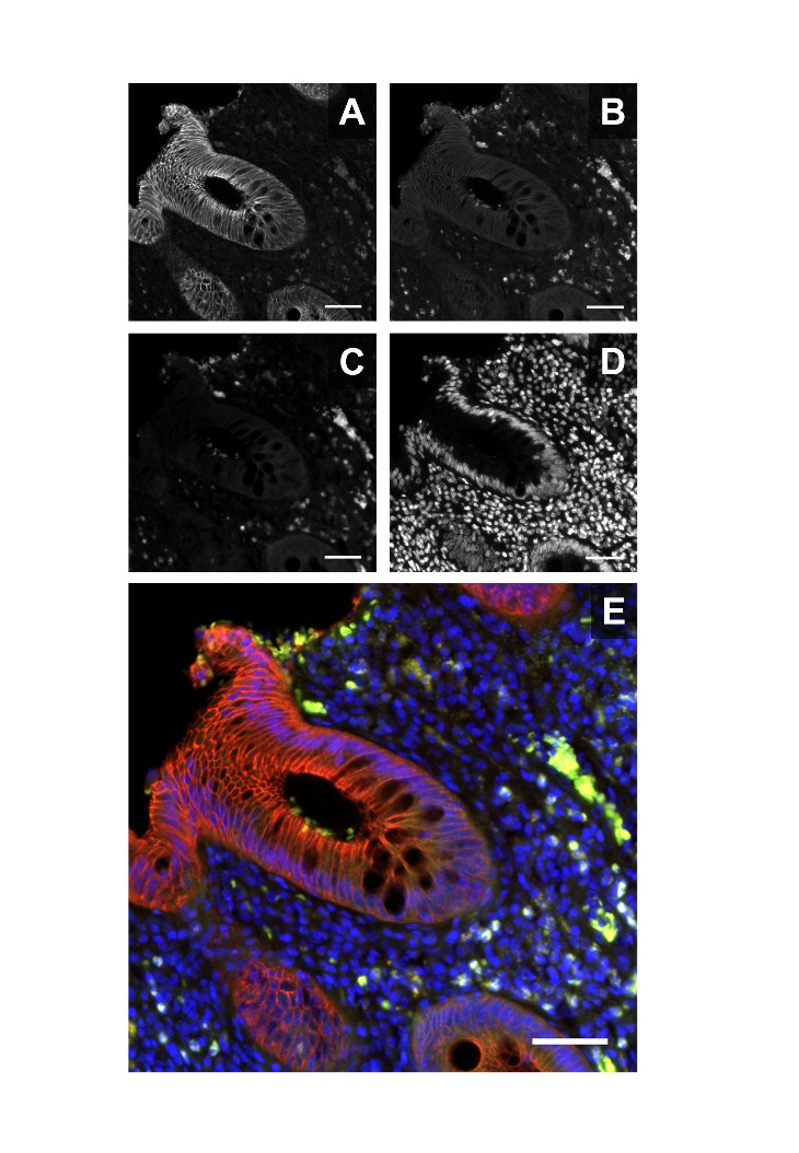
-embedded tissues is a frequently used method to answer research questions or even detect the abundance of a certain protein for diagnostic use. However, the signal originating from specific antibody-staining might be distorted by lt autofluorescence (AF) of the assessed tissue. Although the AF phenomenon is well known, its presence is often neglected by insufficient staining controls. In this study, we describe the existence of cellular AF in paraffin-embedded healthy and inflamed human and murine colonic tissues and present ways to reduce AF. The AF signal is detectable at emission spectra from 425 nm–738 nm, upon excitation from 403.6–638.7 nm and appears more pronounced in inflamed tissues. Most signals are located subepithelially in the tissue and in blood vessels. Previous studies have shown that the AF signals are caused by lipofuscin, which accumulates in lamina propria lt immune cells. In murine small intestine AF signals are present in granules in the Paneth cell zone. For alleviation of the AF signal, sudan black b (SBB) or copper lt sulfate was used. Incubation of the tissue slices with either one of the substances reduced AF. In conclusion, AF appears as an intrinsic biomarker for colonic inflammation. The dominant existence of AF in immune cells of IBD tissue elucidates the importance of negative controls and the limitation of IF staining for potential diagnostic purposes.


Figure: E-cadherin IF staining and autofluorescence in human colonic tissue. E-cadherin IF is detectable in the colonic epithelium in the far-red (650nm) excitation spectrum when a Cy5-labeled secondary antibody is used (A). Excitation of the same specimen at 592nm results in a faint E-cadherin signal, while a stronger signal is detectable in the subepthelial space (B). At 488nm excitation, the E-cadherin signal is absent, while the subepithelial located signal is still detectable (C). DAPI was used for cellular counterstaining (D). The overlay image shows the colocalization of the unspecific autofluorescence signals from the 592nm and 488nm channels and the specific E-cadherin and DAPI signals (E). All images were recorded from a single staining of one specimen. Scale bar: 50μm.


Suchen
Back to content | Back to main menu